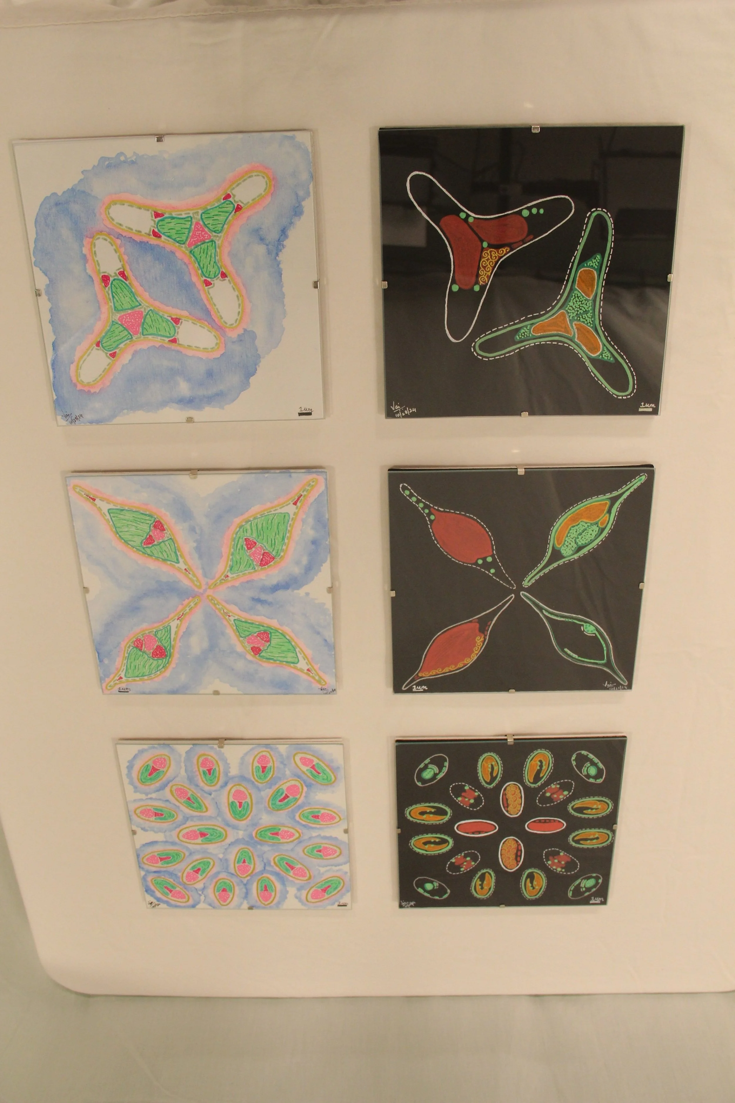
Morphology and Skeleton
Year : 2024
Media : Water and acrylic colours
Price per piece : $50
Please include the name of the piece in the Venmo info
Morphology and Skeleton is an interpretive illustration showcasing the various morphotypes of the model diatom studied here, specifically Phaeodactylum tricornutum. This diatom belongs to a distinct class of unicellular microalgae known for their unique properties. The work comprises a six-piece illustration, with each canvas measuring 8 X 8 inches. The three white canvases depict the morphological variations of the diatom observed under a bright field microscope at different growth phases, highlighting the differences in internal organelles. A bright-field microscope uses light to illuminate objects and helps view magnified images (100X). In contrast, the three black canvases illustrate how cellular organelles appear when stained and viewed under a fluorescence microscope. Fluorescence refers to the property of certain pigments that emit visible light after absorbing non-visible radiation. The microscope that exploits this property to visualize and magnify objects is called a fluorescence microscope. Similar acrylic pigments have been utilized to replicate the appearance of the pigments used in microscopy, providing viewers with an experience akin to that of a fluorescence microscope. The contrasting canvases create a clear distinction between the experiences of bright-field and fluorescence microscopy for the viewers.