Canvas Illustrations
Diversity
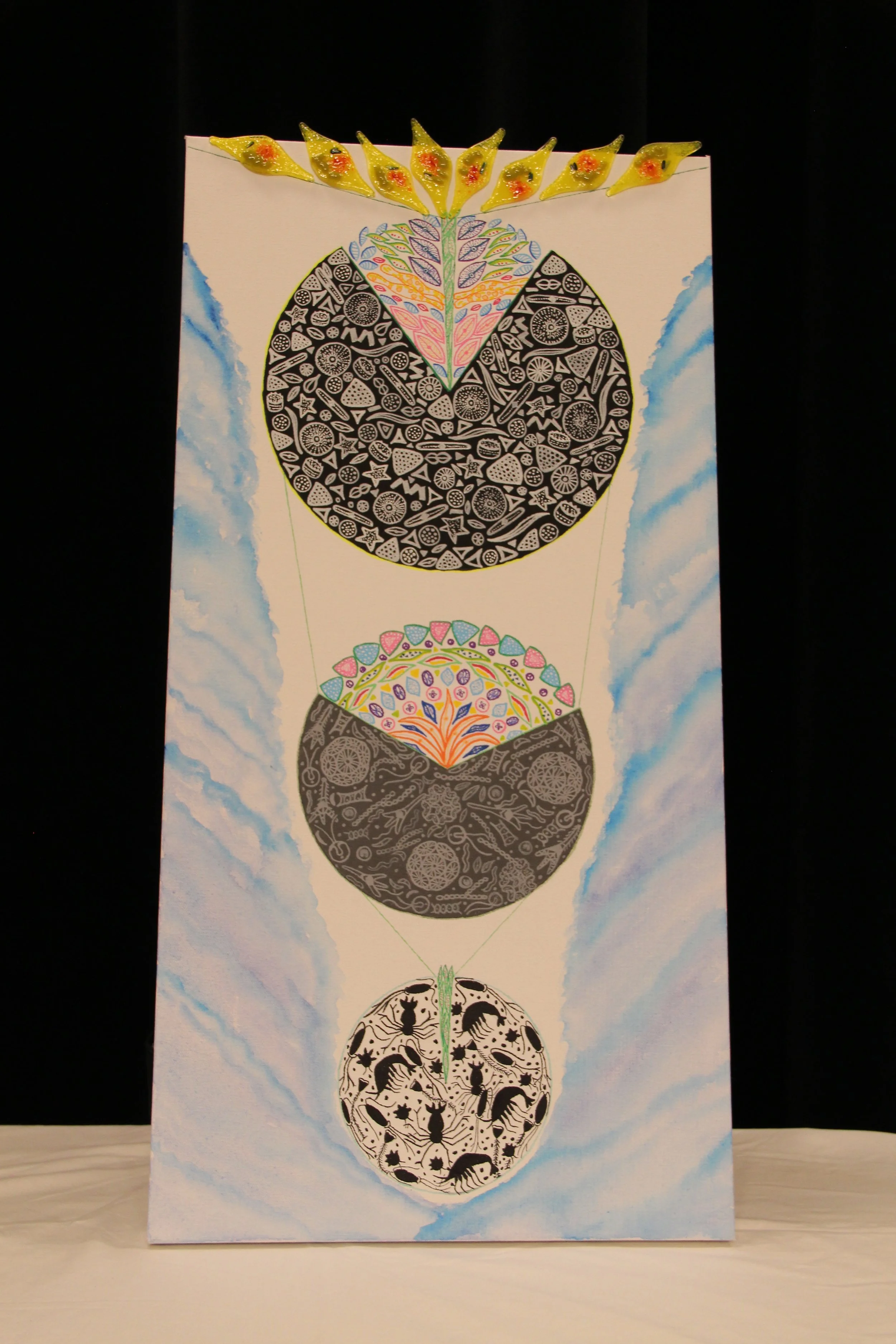
Diversity showcases the vast array of microscopic plankton found in the ocean. These tiny organisms drift along with ocean currents and tides, playing a crucial role in the marine food chain. Plankton primarily consists of microscopic plants, known as “phytoplankton,” and microscopic animals, referred to as “zooplankton.” This piece measures 16 X 40 inches and features three pie charts that emphasize the biodiversity observed in the ocean. Each chart represents biomass abundance, which is the amount of organic material, such as plants and animals, in a defined region. The pie charts are divided into focus groups (colored) and non-focus groups (black, grey, and white). The bottom pie chart illustrates the differences in biomass abundance between phytoplankton (colored) and zooplankton (black). The middle pie chart highlights the diversity within the phytoplankton group, specifically focusing on the diatom group of algae (colored). Finally, the topmost pie chart delves into the diversity within the diatom group, with a particular emphasis on pennate diatoms (colored), which contain the model diatom for this study.
Under the electron microscope
An interpretive illustration, Under the Electron Microscope, showcases the imaging of the model diatom, Phaeodactylum tricornutum (PT). It is an 18 x 24 canvas illustration that depicts the lead author's view of the diatom at 50,000 times magnification. Diatoms are a special class of algae with silica cell walls (commonly known as quartz, a glass-like substance), which make up 40% of the phytoplankton abundance in the ocean. The lead author has demonstrated different types of electron microscopy imaging techniques, including scanning electron microscopy (SEM) and transmission electron microscopy (TEM). Electron microscopy uses electron beams to create magnified images. an electron is a negatively charged particle that orbits a very small particle called an atom, similar to how planets orbit the sun. SEM helps image the exterior surfaces of the sample, whereas TEM creates images by passing electrons through the samples. In our interpretive illustration, the lead author utilized SEM in the scientific study and interpreted the TEM for the interior organelle structure (the colored internal organs of the diatom) of the PT based on the current understanding of scientific literature. Zentangle art was used to create the abstract shapes of the organelles present in the diatom.
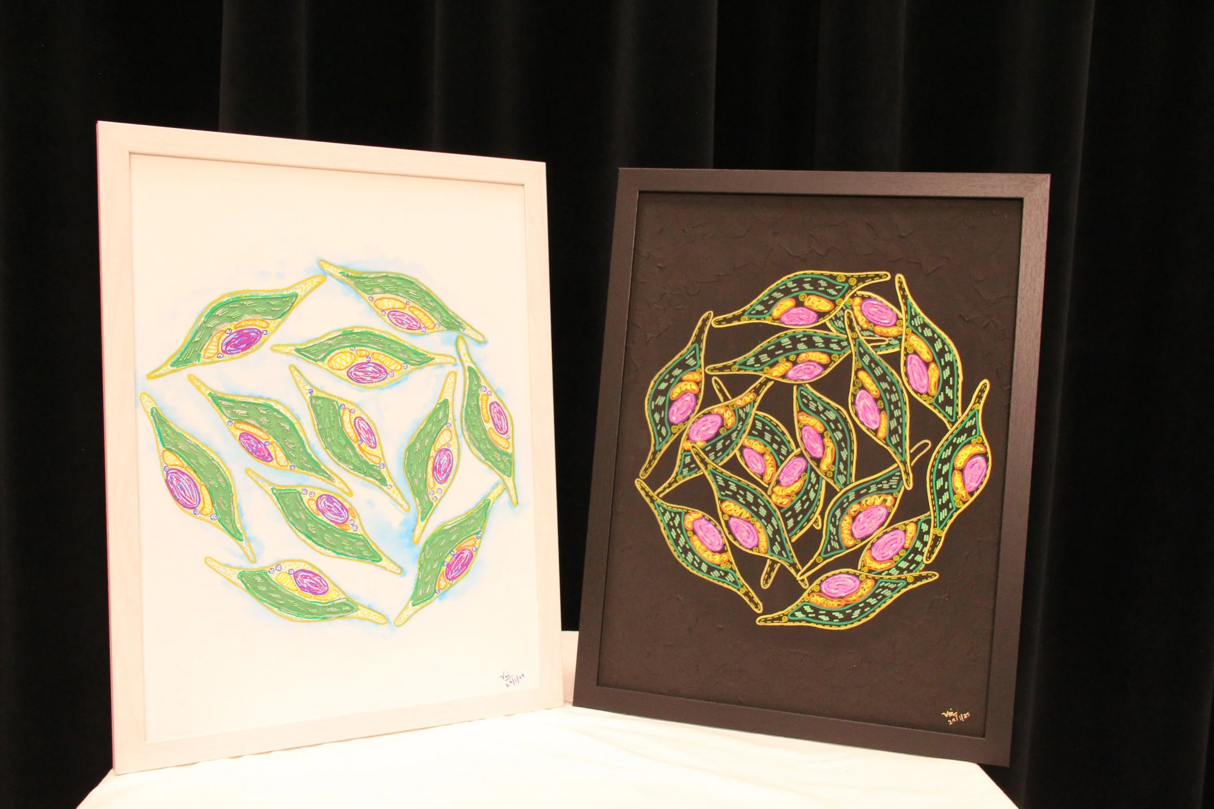
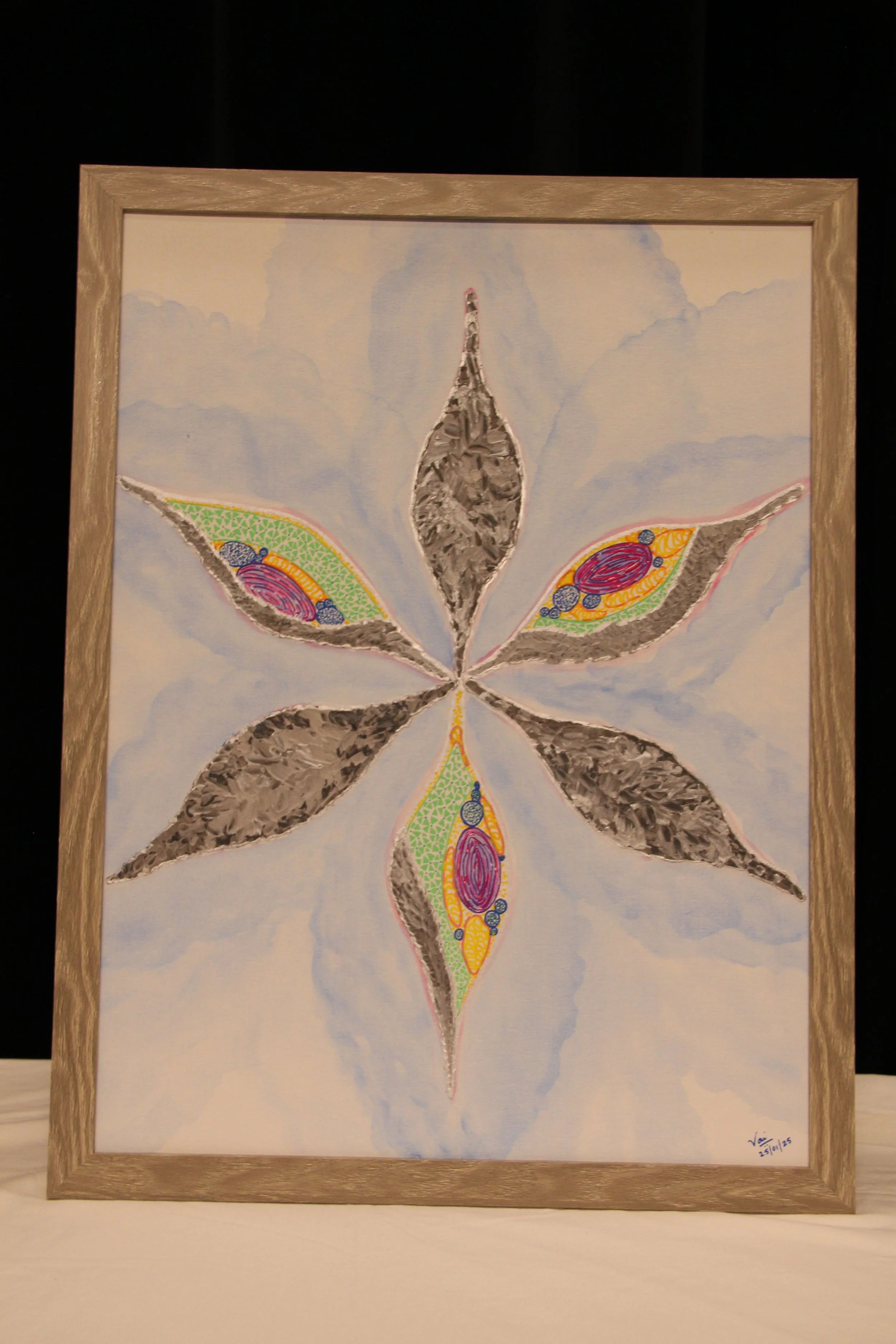
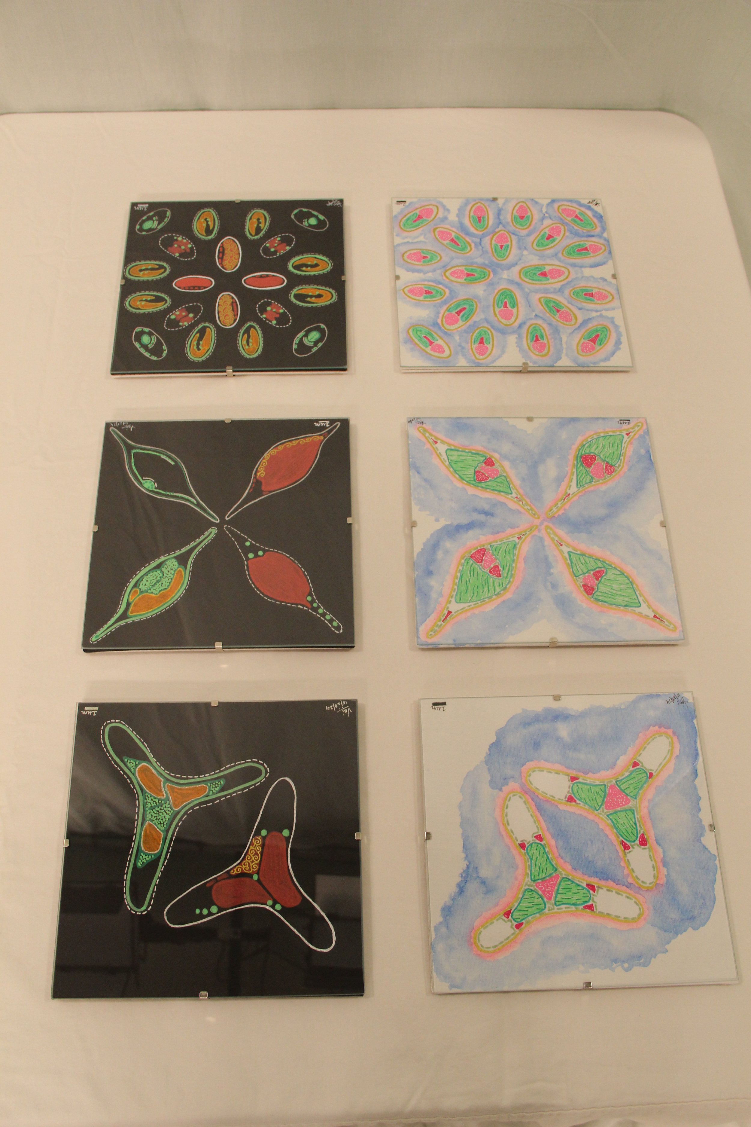
Morphology and Skeleton
Morphology and Skeleton is an interpretive illustration showcasing the various morphotypes of the model diatom studied here, specifically Phaeodactylum tricornutum. This diatom belongs to a distinct class of unicellular microalgae known for their unique properties. The work comprises a six-piece illustration, with each canvas measuring 8 X 8 inches. The three white canvases depict the morphological variations of the diatom observed under a bright field microscope at different growth phases, highlighting the differences in internal organelles. A bright-field microscope uses light to illuminate objects and helps view magnified images (100X). In contrast, the three black canvases illustrate how cellular organelles appear when stained and viewed under a fluorescence microscope. Fluorescence refers to the property of certain pigments that emit visible light after absorbing non-visible radiation. The microscope that exploits this property to visualize and magnify objects is called a fluorescence microscope. Similar acrylic pigments have been utilized to replicate the appearance of the pigments used in microscopy, providing viewers with an experience akin to that of a fluorescence microscope. The contrasting canvases create a clear distinction between the experiences of bright-field and fluorescence microscopy for the viewers.
Light and dark activities of the diatom
This two-piece canvas illustration captures the contrasting activities that take place within diatom cells. Each canvas measures 18 X 24 inches and features a mixed media illustration employing the impasto acrylic technique to emphasize the organelles that are active during both the light and dark phases of diatom activity. In the ocean, diatoms undergo diel regulation, a term that describes the light and dark phases experienced by diatom over a 24-hour cycle. Vaishnavi conducted measurements of volatile organic compound (VOCs) production throughout this diel regulation in diatoms, presenting two key findings (REF). First, it was found that VOCs are produced in significant amounts during the day, primarily through chlorophyll, represented by the green elevated region of the diatom. Chlorophyll is a pigment that enables diatoms to generate their energy source, which is akin to how plants produce food. This process, known as carbon fixation, occurs via photosynthesis, allowing diatoms to create food using light and carbon dioxide. Second, during the dark phase, diatoms engage in reproduction, depicted in purple. Additionally, diatoms continue to produce VOCs in the dark utilizing mitochondria, represented by the yellow elevated organelle. Mitochondria are essential for energy production, similar to how a person sweats during exercise when muscles are active. In this illustration, the lead author simplifies the findings regarding VOC production in diatoms.